Supplement to CRST Global (2026 Vol. 21 No. 2) · Sponsored by SIFI S.p.A.
Harnessing Spherical Aberration for Functional Vision
Leading surgeons share their clinical experiences from the SIFI IOL Expert Forum held on October 25, 2025—exploring how wavefront-engineered spherical aberration modulation creates a continuous range of functional vision without light splitting.
At a Glance: SIFI IOL Portfolio
- EVOLUX® — Partial DoFi-Enhanced (~1.40–1.50 D) enhanced monofocal with smooth wavefront modulation & minimal dysphotopsias.
- MINI WELL® & MINI WELL® TORIC — Partial DoFi-Extended wavefront EDOF IOL with 3 concentric optical zones.
- MINI WELL PROXA® & TORIC — 5-zone wavefront optic reinforcing near-to-intermediate range.
- WELL FUSION® System — Binocular strategy pairing MINI WELL® (dominant eye) + MINI WELL PROXA® (nondominant eye) for continuous full-range vision.
SIFI & Wavefront Engineering – A Story of Innovation
Shaping the future of vision. Before the industry had a name for it.
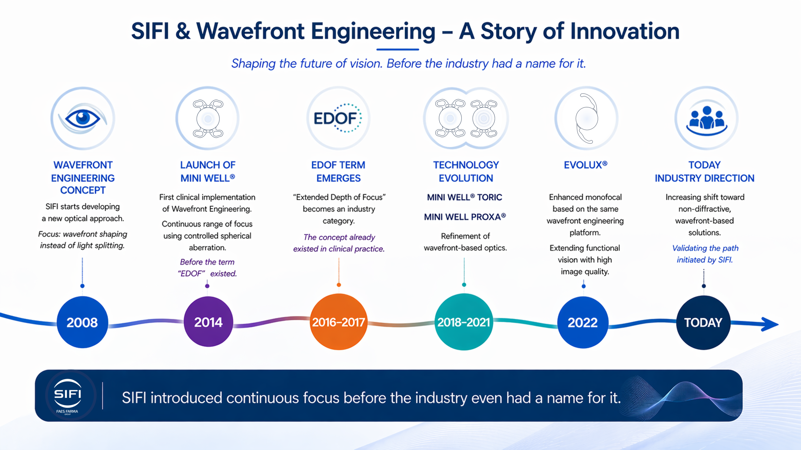
Wavefront Engineering Concept
SIFI starts developing a new optical approach. Focus: wavefront shaping instead of light splitting.
Launch of MINI WELL®
First clinical implementation of Wavefront Engineering. Continuous range of focus using controlled spherical aberration—before the term “EDOF” existed.
EDOF Term Emerges
“Extended Depth of Focus” becomes an industry category. The concept already existed in clinical practice.
Technology Evolution
MINI WELL® TORIC & MINI WELL PROXA®: Refinement of wavefront-based optics.
EVOLUX®
Enhanced monofocal based on the same wavefront engineering platform, extending functional vision with high image quality.
Industry Direction
Increasing shift toward non-diffractive, wavefront-based solutions—validating the path initiated by SIFI.
Executive Overview
Patient demand for spectacle independence across distances continues to grow and shape how ophthalmologists evaluate presbyopia-correcting IOLs. Surgeons and their patients increasingly seek optical designs that extend depth of field while limiting dysphotopsias and preserving contrast sensitivity. To explore the technologies supporting these goals, an international faculty recently convened to discuss the evolution, optical foundation, and clinical results of the SIFI IOL portfolio (SIFI S.p.A.). Their discussion centered on understanding how spherical-aberration modulation, rather than light splitting, can create a controlled range of functional vision, and how these concepts translate into real-world outcomes with EVOLUX®, MINI WELL®, MINI WELL PROXA®, and the WELL FUSION® optical system (a binocular strategy with MINI WELL® implanted in the dominant eye and MINI WELL PROXA® implanted in the nondominant eye). This monograph distills the most clinically relevant takeaways from the faculty’s discussion.
Harnessing Spherical Aberration for Functional Vision
The SIFI Innovation Journey
Maria Cristina Curatolo, PhD, Head of Innovation and Medical Science for SIFI, described the company’s development of presbyopia-correcting optics, which began with its transition from distributor to manufacturer in 2008. During this period, Eng. Curatolo and the research and development team encountered a provisional patent by Junzhong Liang, PhD, that proposed an innovative wavefront engineering technology that induced spherical aberration within specific optic zones. SIFI licensed this concept, which underlies the development of its portfolio.
The launch of the MINI WELL® IOL in 2014 represented the first extended depth-of-focus (EDOF) IOL in the world designed with wavefront technology to feature controlled spherical aberration modulation.1 Over time, the company expanded the patent family to include the MINI WELL PROXA®, MINI WELL TORIC, MINI WELL PROXA® TORIC, and EVOLUX® IOLs.
Foundational Optical Concept: Harnessing Spherical Aberration
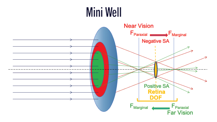
The core principle behind all SIFI presbyopia-correcting IOLs is the controlled modulation of spherical aberration. Vicente J. Camps, PhD, explained that both positive and negative spherical aberration can extend depth of field, but each has limitations when used alone. Positive spherical aberration shifts focus hyperopically and may reduce optical quality, while negative spherical aberration extends depth toward intermediate and near but can compromise distance acuity if over-induced.2 The solution, Dr. Camps noted, is to use carefully calculated combinations of positive and negative aberration within the same optic to achieve depth of field without degrading the retinal image or contrast sensitivity.3
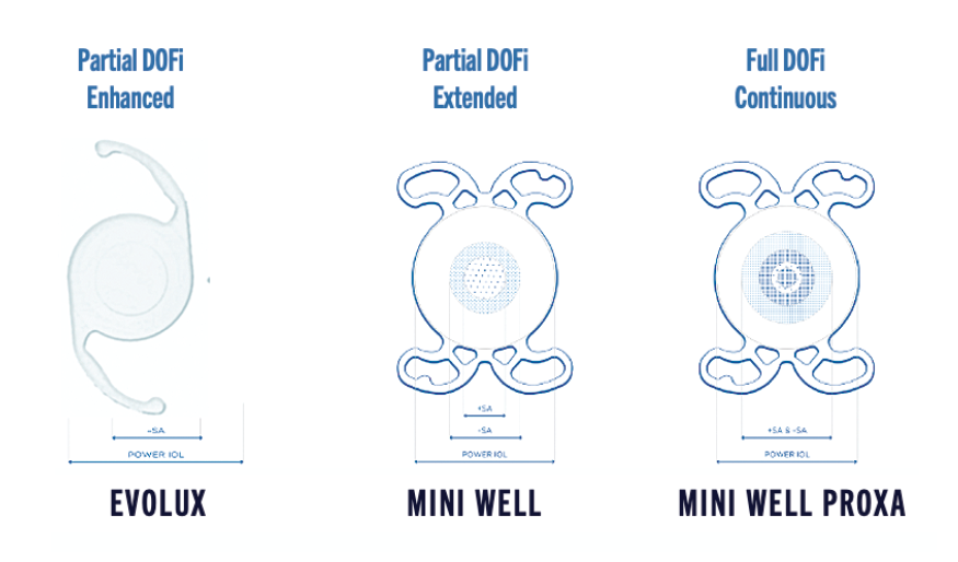
In SIFI’s lenses, these modulations occur in concentric zones, whose number and diameter differ between models. MINI WELL® features three optical zones, including a central refractive zone measuring 3.0 mm to create an extended focal region. MINI WELL PROXA® has five zones and a wider central zone (4.5 mm) to support full-range performance when paired with MINI WELL® in the WELL FUSION® optical system. EVOLUX® features a smoother, low-amplitude modulation profile associated with enhanced intermediate performance and minimal dysphotopsias (Figure 1). The underlying mechanism in all SIFI EDOF lenses involves shifting marginal versus paraxial light to create an elongated, continuous focal distribution—without diffractive steps and without splitting light into discrete foci (Figure 2).
Aberrometric Signatures of the SIFI Portfolio
Prof. Ahmed H. Assaf, MD, PhD, FRCSEd, FEBOS-CR, presented aberrometric results that illustrated the SIFI IOLs’ wavefront characteristics. MINI WELL® demonstrates the characteristic “donut-shaped” signature, with positive/negative modulation across its zones. MINI WELL PROXA® extends these modulations across a wider diameter for a steeper near-intermediate transition. EVOLUX® shows a much smoother, lower-amplitude surface profile, with subtle changes measured in micrometers, yet sufficient to produce clinically meaningful depth of field. These findings align with theoretical studies on the MINI WELL that Dr. Camps and colleagues conducted in Spain.4,5
Rosa Giglio, MD, FEBO, highlighted that these variations occur at scales as small as 10 µm in the central zones of the SIFI IOLs, so that the controlled modification of curvature results in measurable gains in visual performance. For example, although pupil size influences which zones contribute most strongly to image formation, because the spherical-aberration modulation produces a continuous axial intensity profile that creates smooth defocus curves, SIFI IOLs perform consistently across lighting conditions.
Optical and Functional Advantages Compared With Other Technologies
Light Utilization and Contrast Preservation
A central differentiator of SIFI’s spherical-aberration–modulated optics is their nondiffractive architecture. Diffractive multifocal IOLs redistribute incoming light into multiple discrete focal points, inherently reducing retinal illumination. SIFI lenses maintain a single, elongated focal region. This means no energy is “lost” to secondary foci, helping preserve contrast sensitivity, especially under mesopic conditions, where diffractive optics often underperform.6
During the forum, multiple presenters emphasized that contrast preservation is closely linked to light utilization. Mauro Zuppardo, OD, noted that traditional multifocals, even the most advanced trifocals, predictably compromise contrast through light splitting, while EDOF lenses based on spherical aberration avoid this trade-off.7
Halometry and Dysphotopsia Assessment
Dysphotopsias remain an important factor influencing patient satisfaction with presbyopia-correcting IOLs.8 Dr. Zuppardo presented exploratory halometry observations obtained using the Aston Halometer (Aston University) to characterize halo profiles associated with different optical platforms, including MINI WELL®, MINI WELL PROXA®, EVOLUX®, and the WELL FUSION® system.
The analysis was performed on a mathematically normalized angular scale derived from monofocal reference values reported in previously published studies.9–11 Within this modeling framework, halo radius distributions were plotted to enable a structured comparison of the evaluated lens designs.
These observations should be interpreted within the context of applied mathematical normalization, which does not replace prospective clinical evaluation. Dedicated clinical studies specifically designed to assess halometric outcomes would further clarify the real-world performance of the different optical platforms (Figure 3).
These findings were corroborated by patient-reported metrics shared by Prof. Emilio Pedrotti, MD, FEBO (unpublished data). He and his team conducted a randomized clinical trial of 36 patients, 18 implanted bilaterally with the WELL FUSION® system and 18 implanted bilaterally with the FineVision (POD F) diffractive trifocal IOL (BVI Ophthalmic). Using the NEI-RQL questionnaire, the WELL FUSION® cohort reported less perceived glare and higher satisfaction in categories related to clarity and independence. Scores on glare perception were significantly higher (indicating fewer symptoms) for the WELL FUSION® group (77) compared with the trifocal group (38), underscoring the clinical relevance of spherical-aberration–modulated optics in minimizing dysphotopic phenomena.
Structural Surface Analysis
Using an optical profiler with nanometer-scale resolution, Dr. Giglio described how the surface modulations of MINI WELL® and EVOLUX® differ from a monofocal control. When noise and spherical baseline were removed, MINI WELL® revealed a characteristic “hill-and-valley” morphology, although the amplitude of these features measured only a few microns. EVOLUX® exhibited an even smoother central topography with variations of approximately 10 µm—minuscule changes that nonetheless produce measurable depth-of-field enhancements.
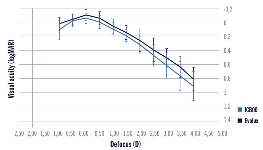
This microstructural analysis echoes Eng. Curatolo’s earlier point that the innovation in SIFI’s optics lies not in dramatic refractive geometry, but in expertly optimized aspheric modulation, where very small changes in the wavefront design lead to significant functional visual benefits. These findings align with comparative defocus curves presented by Dr. Giglio, which showed the binocular performance of the EVOLUX® IOL maintaining a continuous, uninterrupted focal progression from intermediate to distance (Figure 4)—a behavior not consistently observed in diffractive multifocal curves.
Mapping SIFI’s Portfolio to the ESCRS/ASCRS Functional Vision Classification
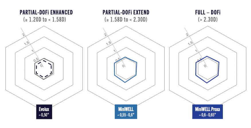
The European Society of Cataract & Refractive Surgeons (ESCRS) and the American Society of Cataract and Refractive Surgery (ASCRS) have recently adopted a function-driven framework for categorizing presbyopia-correcting IOLs.12,13 Instead of classifying lenses by their optical mechanism (eg, diffractive, refractive, nondiffractive EDOF), the new model focuses on clinical performance, using two primary metrics:
- Monocular defocus curves above thresholds of 0.2/0.3 logMAR; and
- Slope between intermediate and near, indicating whether the depth of field is narrow, enhanced, extended, or full range.
Carlos Rocha-de-Lossada, MD, PhD, FEBO, explained that this method reflects how patients experience vision with IOLs, rather than how the optics are engineered. It also better discriminates between lenses with subtle differences in depth of field (DoFi) or functional range.
During the discussion, presenters reviewed monocular and binocular defocus curves across IOL platforms. For example, Giacomo Savini, MD, showed that EVOLUX® achieved a mean DOFi of ~1.40 D, placing it within the Partial DoFi-Enhanced category.
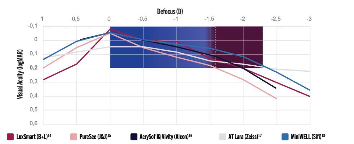
In the Partial DoFi-Extended category, MINI WELL® demonstrated superior intermediate performance compared with LuxSmart (Bausch + Lomb) (Figure 5).14–18 Eng. Curatolo explained that such variation results from SIFI’s carefully optimized coefficients of spherical aberration.
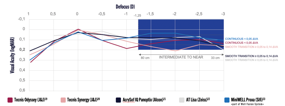
For the FULL DOFi IOL category, MINI WELL PROXA®’s defocus curve aligned with advanced multifocal benchmarks while avoiding their drawbacks related to light-splitting (Figure 6).19–23 When paired with MINI WELL® in the WELL FUSION® system, the binocular curve became continuous, with no gaps between intermediate and near distinction, highlighted during the forum as a hallmark of the WELL FUSION® design.
Why This Matters Clinically
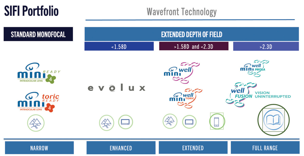
Understanding where each lens falls within functional categories helps surgeons tailor solutions to patients’ expectations. For patients prioritizing image quality and low dysphotopsias, enhanced-range lenses like EVOLUX® offer clarity with expanded intermediate activity. Those seeking broader spectacle independence may benefit from MINI WELL®, while patients demanding seamless full-range functional vision—such as active individuals or those frequently transitioning between focal distances—may be best served by the WELL FUSION® system (Figure 7). The classification also supports more streamlined communication with patients. Rather than describing optical mechanics, surgeons can reference functional scenarios: reading, dashboard viewing, computer work, or nighttime driving.
Clinical Experience: Prof. Assaf’s First 100 Patients Implanted With WELL FUSION®
Professor Ahmed Assaf, MD, PhD, FRCSEd, FEBOS-CR, presented outcomes from his first 100 clinical cases (unpublished) using the WELL FUSION® optical strategy, illustrating how the MINI WELL® and MINI WELL PROXA® IOLs perform together in real-world patients. Prof. Assaf stressed that these are not “mix-and-match” lenses, but a designed pair, identical in material, biocompatibility, and mechanical behavior. Their difference lies solely in their wavefront-engineered profiles. MINI WELL® favors the intermediate-to-distance range, while MINI WELL PROXA® reinforces near, extending the combined binocular range beyond what bilateral MINI WELL® can achieve.
He noted that he typically places MINI WELL® in the “dominant” position that best supports the patient’s functional needs (for example, implanting the MINI WELL PROXA® lens in the second eye, irrespective of dominance, if the patient places more value on near visual performance).
Visual Acuity and Defocus Performance
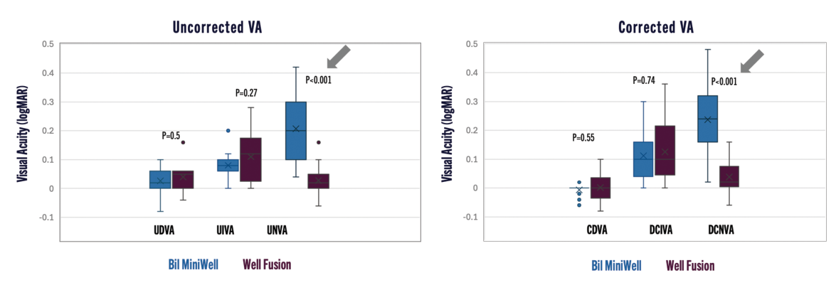
Prof. Assaf’s 100-patient cohort demonstrated strong monocular and binocular visual acuity results 3 months post-surgery (Figure 8). Compared with bilateral MINI WELL® implantation, the WELL FUSION® combination produced lower near UNVA, comparable or improved intermediate vision, and superior binocular distance-corrected near acuity (DCNVA) (all differences reached statistical significance, P<.001).
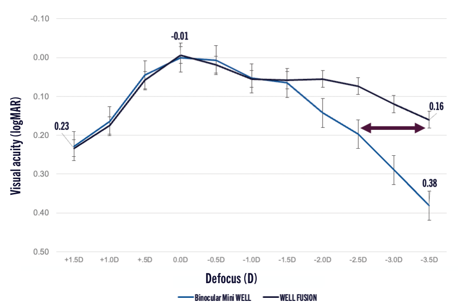
Defocus curve analysis showed that the pairing produces a full-range defocus profile with ΔVA < 0.14 logMAR from distance through near (Figure 9). This was most evident in the MINI WELL PROXA®-enhanced near segment, where visual acuity remained stable across a wider dioptric span compared with binocular MINI WELL®. Prof. Assaf reported that the WELL FUSION® system extended depth of field by approximately 1.00 D compared with bilateral MINI WELL®, aligning with the ESCRS/ASCRS functional-vision classification.
Spectacle Independence and Photic Phenomena
The WELL FUSION® group demonstrated greater spectacle independence than bilateral MINI WELL® recipients, and there were minimal photic disturbances and low reliance on reading glasses across the cohort. Importantly, halo perception did not increase with the addition of MINI WELL PROXA®. Prof. Assaf concluded that WELL FUSION® provides continuous full-range functional vision with minimal dysphotopsias.
Comparative Study: WELL FUSION® vs a Trifocal IOL
In Prof. Pedrotti’s comparison study of the WELL FUSION® system versus the FineVision (POD F) diffractive trifocal IOL (BVI Ophthalmic), described earlier in this monograph, both groups achieved excellent distance vision, but the trifocal recipients showed a slight photopic advantage. WELL FUSION® delivered superior intermediate acuity, comparable near acuity, and a gap-free defocus curve, unlike the stepped profile of the trifocal (36 eyes in both groups; unpublished data). In terms of binocular contrast sensitivity, Prof. Pedrotti noted that the study “confirmed the quality of vision of the WELL FUSION® system,” with better results for WELL FUSION®. Under photopic conditions, the FineVision IOL recorded similar results to the WELL FUSION® system, while under mesopic and scotopic conditions, the WELL FUSION® system showed better contrast sensitivity. Finally, the results obtained on halos confirmed the significantly lower risk of dysphotopsia with the WELL FUSION® system compared to a trifocal IOL and showed no significant difference compared to the gold-standard monofocal IOLs.
Real-World Experience: Dr. Ster’s 100-Patient Analytical Study
Sever Ster, MD, presented outcomes from a 100-patient (200-eye) analytical study (unpublished) on how the WELL FUSION® system delivers a continuous range of functional vision. His cohort reflected typical cataract patients: a mean age of 61.3 years, a balanced gender distribution, axial lengths ranging from 21.02 to 26.01 mm, and no concomitant ocular pathology. Each patient received MINI WELL® in the dominant eye (or its toric version when indicated), and surgeries were performed 2 days to 1 month apart. The refractive target was <0.75 D using the Barrett and Kane formulas, and the study followed patients at 1 day, 1 month, and 3 months after surgery.
Predictability and Stability
The refractive outcomes were highly predictable and stable. By 3 months, 85% of eyes were within ±0.50 D of target, and 95% were within ±0.75 D of target. Dr. Ster reported no cases of IOL decentration or tilt and excellent in-the-bag positioning at all visits.
Visual Acuity and Defocus Performance
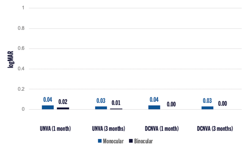
Visual acuity improved progressively from 1 to 3 months across all distances. Mean binocular UDVA and BCDVA remained between 0.0 and 0.1 logMAR. At 60 cm, binocular UIVA reached 0.0 logMAR in 85% of patients at 1 month, increasing to 90% at 3 months, while 95% achieved 0.0 logMAR DCIVA. Near vision at 40 cm and 33 cm (Figure 10) was similarly strong, with 90% achieving 0.0 logMAR binocularly by 3 months, and all patients reaching ≤0.10 logMAR, even under low-light conditions.
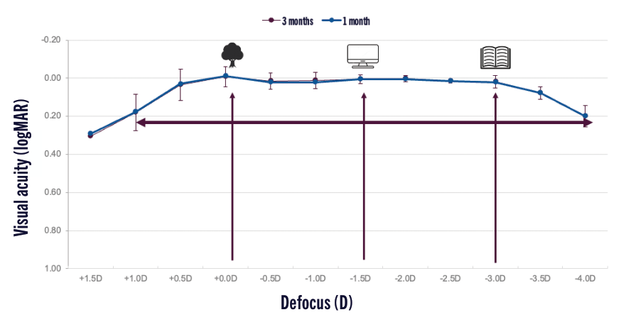
Binocular defocus analysis revealed a smooth, dome-shaped curve spanning approximately 5.00 D at 0.2 logMAR or better (+1.00 to −4.00 D), without gaps between distance, intermediate, and near (Figure 11). Dr. Ster described this as evidence of true continuous vision.
Contrast Sensitivity and Clinical Conclusions
Binocular contrast sensitivity was comparable to monofocal performance and to that of a healthy 20- to 30-year-old reference group, with minimal halo or glare reported. Dr. Ster concluded that the MINI WELL PROXA® combination delivers stable, full-range vision, rapid neuroadaptation, and high visual quality, performing consistently across refractive profiles and remaining suitable for patients with mild retinal pathology.
EVOLUX®: Early Real-World Results and Functional Performance
The EVOLUX® platform is SIFI’s most recent iteration of spherical-aberration–based optics, engineered to provide an enhanced depth of field with low dysphotopsias.24 Its design employs a smoother, lower-amplitude surface modulation than MINI WELL® or MINI WELL PROXA®, producing a controlled depth of field of around 1.50 D.
Giacomo Savini, MD, presented a prospective clinical evaluation designed to determine whether the EVOLUX® IOL meets the criteria for the Partial DoFi-Enhanced category within the ESCRS/ASCRS functional classification of presbyopia-correcting IOLs.25 The study included 32 eyes of 32 patients (mean age 76.5 years) with no intra- or postoperative complications and no keratometric astigmatism ≥0.75 D. A monofocal aspheric IOL (AcrySof SA60WF; Alcon) served as the control group (27 eyes). DOFi was defined as the dioptric range over which mean visual acuity remained 0.2 logMAR or better.
Key Clinical Results
- Mean DOFi for EVOLUX®: 1.42 ±0.56 D, significantly greater than the monofocal control group’s 1.04 ±0.54 D (P = .0272).
- DOFi Distribution: Varied from 0.50 to 2.50 D—with 34% showing <1.20 D and 28% showing >1.58 D, an expected finding for enhanced-range lenses.
- Wavefront Analysis: Demonstrated negligible induced internal spherical aberration (0.00 µm at 3 mm), indicating that EVOLUX® does not significantly modify the eye’s intrinsic aberration profile.
- Visual Disturbances: Patients reported no subjective visual disturbances, and contrast sensitivity remained comparable to monofocal controls under both photopic and mesopic conditions.
Conclusion: Dr. Savini concluded that EVOLUX® qualifies as a Partial DoFi-Enhanced IOL within the ESCRS/ASCRS functional classification. Its greater depth of field, superior intermediate performance, and lack of dysphotopsias differentiate it from a monofocal IOL while avoiding the light-splitting artifacts seen in diffractive designs.
Comparative Evidence: EVOLUX® vs Enhanced Monofocal & EDOF Competitors
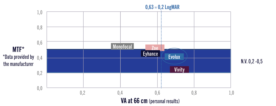
Andrea Bedei, MD, presented visual acuity and defocus comparisons between EVOLUX® and other enhanced or EDOF lenses, including Eyhance (Johnson & Johnson Vision), ZOE (Ophthalmo Pro GmbH), and Clareon® Vivity® (Alcon Laboratories, Inc.). At 66 cm, a critical working distance for intermediate tasks, EVOLUX® consistently showed better acuity than those lenses (Figure 12).
Dr. Bedei also described the outcomes of one patient in whom he’d implanted an EVOLUX® IOL OS following the implantation of an Eyhance IOL OD a few years prior. The patient reported no difference in distance vision between the two eyes (both achieved a postoperative UDVA of 1.00 D), but better intermediate performance in the eye implanted with EVOLUX®. This outcome supports the versatility of EVOLUX® in hybrid implantation strategies.
Patient-Reported Outcomes and Spectacle Independence
Dr. Giglio and her team evaluated visual outcomes and daily task performance after bilateral implantation of two enhanced monofocal IOLs: the ICB00 TECNIS Eyhance (n=28) and SIFI’s EVOLUX® (n=14) (see Figure 4). In addition to evaluating postoperative refractive outcomes, they administered the revised Heidelberg Daily Task Evaluation (DATE) questionnaire, which measures spectacle independence in everyday activities (unpublished data).
They found similar levels of real-world spectacle independence between the IOLs, with patient-reported outcomes aligning with targeted acuities. Dr. Giglio noted that EVOLUX® patients especially appreciated the natural visual quality and minimal halo or glare.
Monocular Implantation and Early Clinical Experience
Dr. Rocha-de-Lossada referenced a published study on the 1-year clinical outcomes of 82 patients (116 eyes) implanted bilaterally with the EVOLUX® IOL.26 By 12 months, the authors reported that 62% of eyes achieved 20/20 or better uncorrected distance visual acuity (UDVA), and 96% achieved at least 20/40. Additionally, 76% of eyes were within ±0.50 D of their postoperative spherical equivalent.
In the forum, he shared two cases of successful monocular implantations of EVOLUX® (an important finding given that many EDOF or multifocal lenses rely on binocular summation to achieve full performance):
Case 1: A female patient presented with aphakia OS due to ocular trauma 30 years prior (CDVA with +12.00-D correction was 0.5 Decimal, equal to 0.3 LogMAR) and complaints of poor vision OD due to cataract (-1.25 -0.25 x 110º). After EVOLUX® implantation in the cataractous eye, the patient’s refraction was +0.5 -0.5 x 137º OD.
Case 2: A male patient presented with a refraction of -0.75 -1.25 x 42º OS due to cataract. The right eye had slight amblyopia and a CDVA of 0.5 LogMAR. After receiving the EVOLUX®, the left eye’s refraction was +0.75 -1.25 x 50º.
In both cases, the EVOLUX® IOL delivered stable acuity and high-quality vision after monocular implantation, making it a viable option when indicated.
Patient Selection, Special Cases, and Practical Recommendations
Patient Selection Guidelines
Throughout the SIFI presentations, the surgeons emphasized that spherical-aberration–based IOLs are generally forgiving and adaptable, but thoughtful preoperative evaluation remains essential. Key preoperative considerations include:
- Pupil Size: Aberration analysis presented by Dr. Pedrotti showed that with a 3-mm pupil, differences between WELL FUSION® and trifocal optics were minimal. However, with 4-mm pupils (representative of mesopic conditions), the WELL FUSION® group demonstrated better root-mean-square (RMS) and Strehl ratio values than the trifocal group, suggesting higher optical quality under low light. This improvement was most pronounced in the dominant MINI WELL® eye and was maintained binocularly. Patients who frequently function in dim environments (driving at night, theater lighting) may benefit from the WELL FUSION® system’s mesopic stability.
- Corneal Aberrations: The speakers agreed that SIFI lenses preserve contrast and tolerate mild corneal irregularity better than diffractive multifocals. However, significant coma or irregular astigmatism can degrade the precision of spherical-aberration–based optics.
- Ocular Surface and Tear Film: As with any premium IOL, an optimized tear film is required to maintain the wavefront quality delivered by the lens.
- Retinal Status: Panelists commented that the MINI WELL®, MINI WELL PROXA®, and EVOLUX® may be used in eyes with mild retinal pathology, where contrast sensitivity could otherwise be a concern.
Post-LASIK, Corneal Pathology, and “Special Case” Considerations
Several presenters discussed how spherical-aberration–engineered optics can behave predictably in eyes that might not be ideal candidates for diffractive multifocals:
- Post-LASIK eyes: Because SIFI lenses do not introduce diffractive steps, they may be more tolerant of corneal higher-order aberrations than diffractive designs. Prof. Assaf’s and Dr. Ster’s experiences indicate that patients tolerate small refractive surprises (±0.50 D) and maintain good distance vision and functional range.
- Eyes with corneal pathology (astigmatism, keratoconus, keratoplasty, Fuchs dystrophy): Salvador García Delpech, MD, PhD, stated that these eyes can accept SIFI optics, with the caveat that “we need to know the lens and know the corneal disease,” because such pathologies will affect the refraction/diffraction of the implanted lens.
- Irregular or mildly aberrated corneas (coma, trefoil, spherical aberration): Wavefront-based designs maintain more stable image quality than multifocal optics when corneal aberrations are modest.27 However, extreme irregularity remains a relative contraindication.
- Pseudophakic surprises: Prof. Assaf described how MINI WELL PROXA®/MINI WELL® recipients can tolerate a mild myopic or hyperopic offset (~0.50 D) without loss of functional acuity, due to the lens’ extended focus zone. When residual error exceeds this range, standard enhancement strategies apply.
Monograph Conclusion
The faculty’s combined optical analyses, aberrometric measurements, functional classifications, and real-world clinical results illustrate the strength of SIFI’s spherical-aberration–modulation approach. The EVOLUX®, MINI WELL®, and MINI WELL PROXA® platforms consistently deliver high-quality vision across functional ranges, low dysphotopsias, strong contrast sensitivity, predictable refractive outcomes, and flexibility for diverse patient scenarios. Together, these attributes reinforce SIFI’s contribution to modern presbyopia-correcting IOL strategies—a customizable continuum of solutions grounded in scientifically optimized optics and validated by extensive surgeon experience.
Faculty Contributors
International faculty participating in the SIFI IOL Expert Forum held on October 25, 2025. All participants are paid speakers/consultants of SIFI S.p.A.

Ahmed H. Assaf
MD, PhD, FRCSEd, FEBOS-CR
Ain Shams University & Assaf Clinic, Cairo, Egypt

Andrea Bedei
MD
Casa di Cura San Camillo, Lucca, Italy

Vicente J. Camps Sanchis
PhD
University of Alicante, Alicante, Spain

Rosa Giglio
MD, FEBO
Eye Clinic, University of Trieste, Trieste, Italy

Salvador García Delpech
MD, PhD
Co-director Aiken Clinic, Valencia, Spain

Emilio Pedrotti
MD, FEBO
Ophthalmology Clinic, University of Verona, Italy

Carlos Rocha-de-Lossada
MD, PhD, FEBO
Qvision, Hospital VITHAS Almería & Málaga, Spain

Giacomo Savini
MD
Studio Oculistico d’Azeglio & IRCCS Bietti, Rome, Italy

Sever Ster
MD
Medical Director, Imagister clinic, Satu-Mare, Romania

Mauro Zuppardo
OD
Department of Science, Roma Tre University, Rome, Italy
References (1–27)
- Bellucci R, Curatolo MC. A new extended depth of focus intraocular lens based on spherical aberration. J Refract Surg. 2017;33(6):389-394.
- Holladay JT, Kozhaya K, Koch DD, et al. Impact of spherical aberration on visual quality and depth of focus. J Cataract Refract Surg. 2024;50(6):651–652.
- Rocha KM, Vabre L, Chateau N, et al. Expanding depth of focus by modifying higher-order aberrations induced by an adaptive optics visual simulator. J Cataract Refract Surg. 2009;35(11):1885-1892.
- Miret JJ, Camps VJ, García C, et al. Analysis of the effect of pupil size and corneal aberration on the optical performance of premium intraocular lenses. J Clin Med. 2025;14:5336.
- Miret JJ, Camps VJ, García C, et al. Optical performance of RayOne EMV and Tecnis Synergy under varying pupil sizes and corneal aberrations. J Clin Med. 2026;15:1095.
- Li J, Sun B, Zhang Y, et al. Comparative efficacy and safety of all kinds of intraocular lenses in presbyopia-correcting cataract surgery: a systematic review and meta-analysis. BMC Ophthalmol. 2024;24(172).
- Camps VJ, Miret JJ, Caballero MT, et al. Multifocal intraocular lenses: basic principles. In: Alio JL, Pikkel J, eds. Multifocal Intraocular Lenses: The Art and the Practice. 2nd ed. Switzerland: Springer;2019:31-42.
- Pusnik A, Petrovski G, Lumi X. Dysphotopsias or unwanted visual phenomena after cataract surgery. Life. 2022;13(1):53.
- Buckhurst PJ, Naroo SA, Davies LN, et al. Tablet app halometer for the assessment of dysphotopsia. J Cataract Refract Surg. 2015;41(11):2424-2429.
- Zeilinger J, Kronschläger M, Schlatter A, et al. Comparing an advanced monofocal with a non-diffractive extended depth of focus intraocular lens using a mini monovision approach. Am J Ophthalmol. 2025;271:86-95.
- Hienert J, Stjepanek K, Hirnschall N, et al. Visual performance of two diffractive trifocal intraocular lenses: a randomized trial. J Refract Surg. 2021;37(7):460-465.
- Ribiero F, Dick HB, Kohnen T, et al. Evidence-based functional classification of simultaneous vision intraocular lenses: seeking a global consensus by the ESCRS Functional Vision Working Group. J Cataract Refract Surg. 2024;50(8):794-798.
- Fernández J, Koch DD, Barrett GD, et al; Functional Vision Working Group. Global consensus on the evidence-based functional classification of simultaneous vision IOLs. J Cataract Refract Surg. 2026;52(3):219-222.
- Campos N, Louriero T, Rodrigues-Barros S, et al. Preliminary clinical outcomes of a new enhanced depth of focus intraocular lens. Clin Ophthalmol. 2021:15 4801–4807.
- Corbett D, Black D, Roberts TV, et al. Quality of vision clinical outcomes for a new fully-refractive extended depth of focus Intraocular Lens. Eye (Lond). 2024;38(Suppl 1):9–14.
- www.myalcon.com
- Song JE, Han SY, Khoramnia R, et al. Clinical outcomes of combined implantation of an EDoF IOL and a trifocal IOL in a Korean Population. J Ophthalmol. 2021;7:2021:9034258.
- Greve D, Bertelmann E, Pilger D, et al. Visual outcome and optical quality of a wavefront-engineered extended depth-of-focus intraocular lens. J Cataract Refract Surg. 2021;47(9):1139-1146.
- Waring G. New full visual range IOL delivers for patients: examining the TECNIS Odyssey intraocular lens. Presbyopia Physician. June 3, 2024. Accessed January 29, 2026.
- Kim J, Kim TI, Seo KY, et al. A comparative study of two presbyopia-correcting intraocular lenses combining bifocal and extended depth-of-focus profiles. Int Ophthalmol. 2024;44(1):54.
- Shin KY, Lim DH, Chung TY. Clinical outcomes after bilateral implantation of trifocal diffractive intraocular lenses and extended depth of focus intraocular lenses. J Clin Med. 2022;11(19):5729.
- Zhu M, Fan W, Zhang G. Stereopsis and visual acuity: bilateral trifocal versus blended extended depth of focus and diffractive bifocal intraocular lenses. Front Med (Lausanne). 2022;9:1042101.
- Mastropasqua L, Pedrotti E, Ruggieri ML, et al. Two-surgeon, two-center evaluation of a new combined EDOF intraocular lens approach. J Cataract Refract Surg. 2023;49(5):512-517.
- Cano-Ortiz A, Sanchez-Ventosa A, Gonzales-Cruces T, et al. Evaluation of EVOLUX®™ intraocular lenses in cataract surgery: clinical outcomes and patient satisfaction. J Clin Med. 2024;13(23):7404.
- Savini G, Galzignato A, Coutinho CP, et al. Functional classification of a new IOL into the category of partial depth of field-enhanced IOLs according to the ESCRS criteria. J Refract Surg. 2026;42(4):e359-e366.
- Gabric K, Jagic M, Gabric I, et al. 12-month clinical outcomes of the EVOLUX®™ extended monofocal intraocular lens following cataract surgery. BMC Ophthalmol. 2025;25:524.
- Miret JJ, Camps VJ, Garcia C, et al. Analysis of the optical performance of intraocular lenses using profilometric measurements. Graefes Arch Clin Exp Ophthalmol. 2024;263:451–465.